

Normal mastication in the horse is performed by the cheek teeth, which consist of three premolars and three molars in each quadrant of the mouth. They can be numbered using the Triadan numbering system (06 to 11). The occlusal surfaces of the cheek teeth are tightly pressed together by the opposing angulation of the 06s, 10s and 11s so they function as a single grinding unit (Collins and Dixon, 2005; Dixon et al, 2008; 2014).
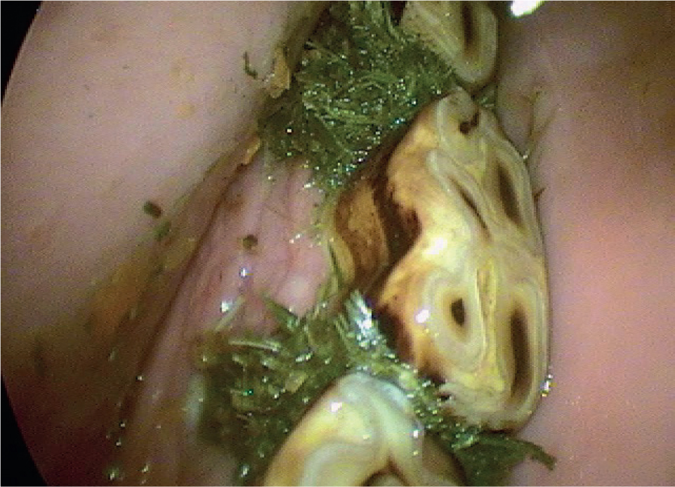
The presence of any space between the cheek teeth is abnormal and is known as a diastema (plural, diastemata). Once a space is present, food can get trapped and impacted between the teeth (Figure 1). Bacteria then proliferate in the decaying food causing gingivitis (gum disease) and the breakdown of the periodontal ligament (periodontitis). Periodontitis can cause great pain to affected horses, particularly when chewing long fibre, making cheek teeth diastema one of the most painful equine disorders (Collins and Dixon, 2005; Dixon et al, 2008; Dixon et al, 2014).
Types of diastemata
There are two different types of diastemata classified. The first is a ‘valve’ type, where the gap at the occlusal surface is narrower than that at the level of the gum. This type is more likely to trap large amounts of food and lead to severe periodontitis. The second type is an ‘open’ type, where the gap between the teeth is the same width from occlusal surface to gingiva (Collins and Dixon, 2005).
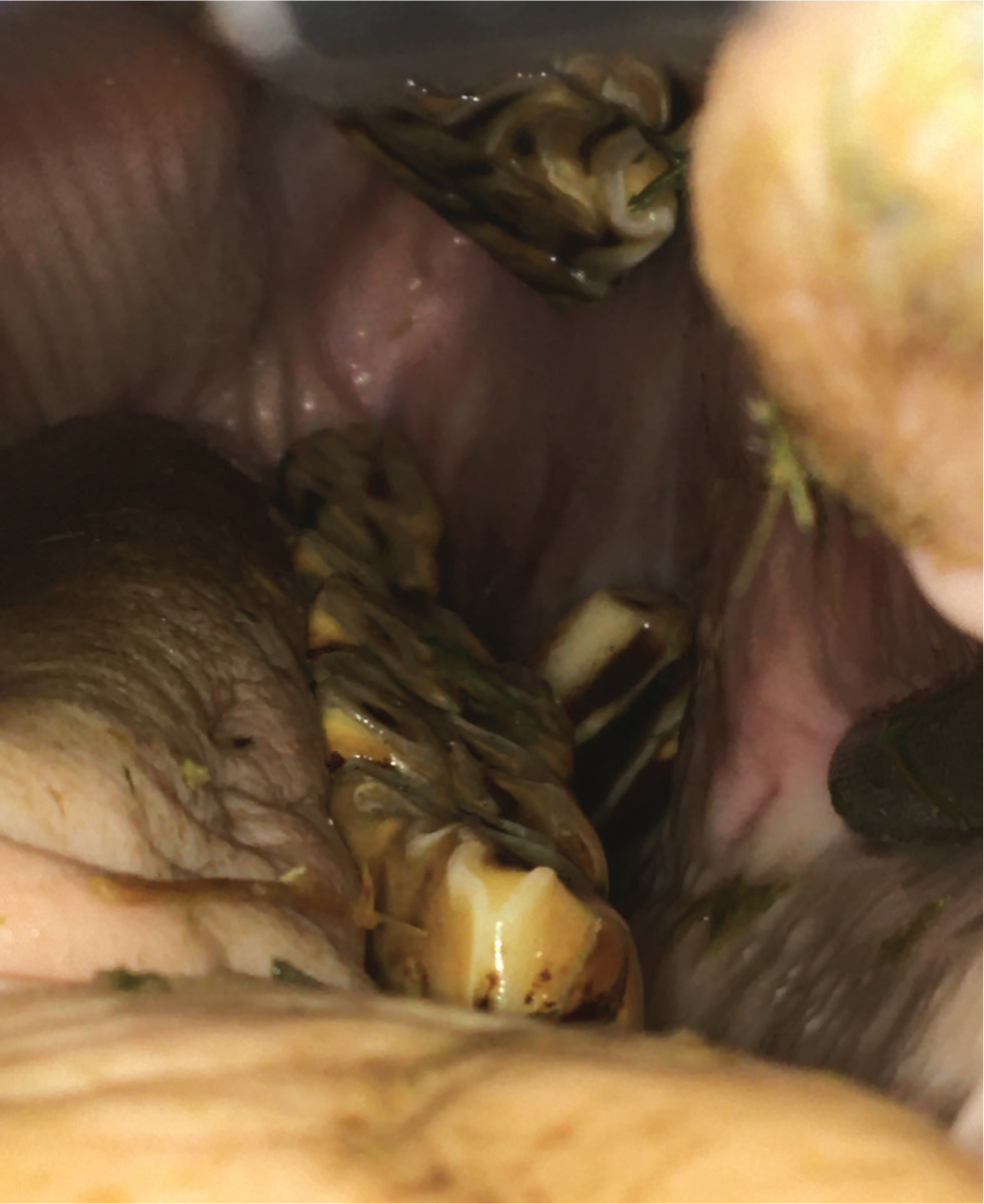
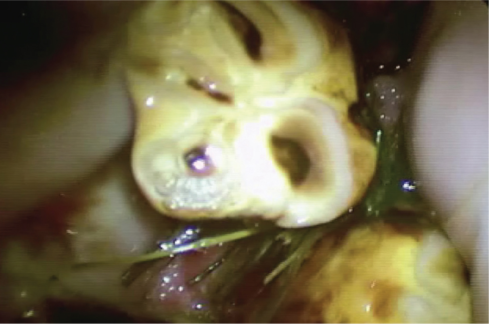
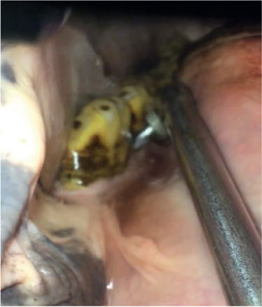
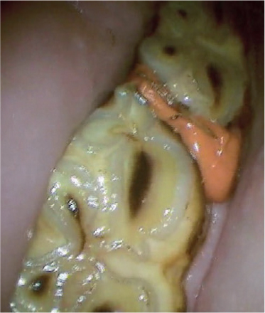
Developmental or primary diastema occur when there is insufficient angulation of the cheek teeth, so they are not tightly compressed together. Developmental diastema may also occur if the cheek teeth have developed too far apart. As this condition is developmental in nature, it affects younger horses as they are developing a mature mouth, typically from 3.5–6 years old (Dixon et al, 2008). Secondary diastema may occur as a result of an extra (supernummerary) cheek tooth within the mouth, that does not fit together well with the existing teeth, leaving an abnormal space between them. Buccal or palatal/lingual displacements of cheek teeth, or the rotation of one of the cheek teeth will also lead to abnormally shaped interdental spaces between teeth. The protruding portion of the displaced tooth may then funnel food into this gap (Figures 2 and 3). Large overgrowths of a cheek tooth can cause the cheek tooth to be displaced and a larger gap to be present.
Secondary diastema can affect horses of all ages (Dixon et al, 2008). Senile diastemata can be present in geriatric animals, as they lose angulation of their cheek teeth and the teeth themselves become narrower towards the roots.
The true incidence of diastemata is unknown, with various studies showing a prevalence of between 29 and 60%, that tends to increase with age (Baker, 1970; du Toit, 2012). A UK study in a first opinion practice showed that 49.9% of horses having dental examinations had diastemata, with 40% of these diastemata being associated with periodontal disease (Walker et al, 2012).
Clinical signs and consequences
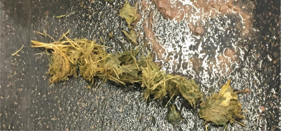
The most common sign of diastemata is ‘quidding’ (dropping balls of semi-masticated food) (Figure 4). Affected horses typically quid their hay or haylage, but eat concentrate feeds normally. Weight loss may also occur if the associated gum and periodontal disease are severe, or if the disorder has been present for some time. Another common sign with diastemata is halitosis, as a result of the decaying food trapped between the teeth. Some affected horses may be inconsistent on the bridle or headshake when ridden (Collins and Dixon, 2005).
In very severe and longstanding cases, the periodontal disease may cause periapical infection or loosening of the teeth. The supporting bones may develop osteomyelitis, which may ultimately have to result in euthanasia. Orosinus fistulation may result from severe periodontal disease affecting the caudal upper cheek teeth, or oronasal fistulation if the more mesial cheek teeth are involved (Hawkes et al, 2008).
Diagnosis
The most common interdental spaces where diastemata develop are the mandibular 09/10s and 10/11s. Diastemata are best diagnosed by a detailed oral examination, using a headlight and dental mirror or oral endoscope. This is best performed with sedation, as affected horses are typically in pain and the most common sites are between the caudal (back) mandibular cheek teeth, where visualisation is difficult because of the large bulk of the tongue. Initial diagnosis should include the site of diastema and its type (‘open’ or ‘valve’).
The next stage in diagnosing diastema is the removal of food from between the teeth and the periodontal pocket. This is extremely painful and should only be carried out under sedation, preferably topical anaesthesia. Removal of the food is both diagnostic and therapeutic. Food can be removed using a combination of instruments including picks, forceps, Hedstrom files and water lavage (Figure 5). An attempt should be made to remove every visible piece of food from between the teeth. The depth of each periodontal pocket should be recorded and teeth adjacent to deep periodontal pockets should be checked for loosening. Radiography is indicated in developmental cases, or if there is suspicion of apical disease, movement in the tooth or infection of the surrounding bone or sinuses.
Treatment options
Fully examining the diastema and flushing all the food material from a periodontal pocket can be considered the first step in treatment. It is common for differing types of diastemata to be present in the mouth and, as such, each should be treated differently. High quality routine odontoplasty should be performed, including removal of exaggerated transverse ridges and the shaping of the edges of displaced teeth. Often, the combination of food removal and good quality dentistry is sufficient to treat a high number most of horses. Removing teeth from occlusion, reducing transverse ridges, extracting displaced cheek teeth, flushing food material and packing periodontal pockets have all been proposed as treatments for equine diastemata (Collins and Dixon, 2005; du Toit, 2012).
The diet in moderate to severely affected horses should be managed. Long fibre (hay or haylage) and even short chopped fibre should be avoided and replaced with either grass, or grass replacement pellets.
Diastema packing
Some clinicians also prevent re-accumulation of food in the periodontal pocket beneath the gum, by filling them with a variety of materials (Townsend, 2015). The most common of these is a vinyl polysiloxane putty (dental impression compound) which sets to a rubber consistency (Figure 6). Care should be taken that all food is removed before placing this into a periodontal pocket, as infection can occur beneath it and lead to continuous pain when chewing. Research has given cause for caution when using packing materials, as they may be toxic to the periodontal tissues, although this was only in an in-vitro situation (Ringeisen et al, 2018).
More permanent packing or ‘bridging’ has been proposed to be effective in selected cases. It has previously been used to treat orosinus fistulae where a diastema is the primary cause (Hawkes et al, 2008). The material used is normally a hard resin composite material, which must either be chemically bonded to the adjacent teeth or mechanically retained by shaping the interdental space to prevent loss of the material. Owing to the hard nature of the material, if it rotates or protrudes from the interdental space; it may cause trauma to the oral soft tissues.
For valve type diastemata, widening with a mechanised burr can be an effective treatment in carefully selected cases (Dixon et al, 2008; 2014). During this procedure, the occlusal portion of the diastema is widened to convert it to an open diastema and prevent food from becoming trapped so readily (Figure 7). This treatment should be performed in a clinical environment, under heavy sedation, because of the risks of permanently damaging the adjacent teeth. A diastema should be widened according to the proximity of the pulp position to the interdental space; in most cases it is preferable to remove more of the mesial portion of the distal tooth (Bettiol and Dixon, 2011).

Extracting the teeth is also a treatment option, particularly for displaced, rotated or supernumerary cheek teeth. In cases with extremely deep periodontal pockets, which are unable to be cleaned effectively, extraction should be considered at a relatively early stage before infection develops in the surrounding osseous structures. Extraction can also assist with treatment of multiple diastemata cases, particularly if the worst diastema are around one tooth. The associated dental drift can allow the remainder of the teeth to space themselves out, although this should be balanced with the other long-term consequences of extraction, such as overgrowth formation (Townsend et al, 2008; Vlaminck et al, 2008).
Prognosis
If diastemata are adjacent to displaced, rotated or supernumerary cheek teeth and are treated before secondary structures are involved, these horses can have a good prognosis. Horses with multiple diastemata can be managed throughout the winter months by repeated flushing of periodontal pockets, at 6–8 weekly intervals. Young horses with developmental diastemata may have a good long-term outlook, particularly if they have good angulation of their cheek teeth, as these may drift together in the long term, with increased eruption. These horses may need intensive management for a number of years, but then they may improve.
KEY POINTS
- Diastemata are a painful condition and the most common cause of quidding.
- Removal of food material and palliative rasping will improve most horses.
- Each diastema is slightly different and should be treated individually.